
Hilum in human anatomy refers to the depression where structures such as blood vessels and nerves enter an organ. The structures contributing to hilar shadows in a Chest X-ray are:
- Major: Pulmonary artery and veins
- Minor: Fat, Lymph nodes and Bronchial walls
Normal Hilum:
- Position: Left hilum is slightly higher than the right hilum
- Shape: Concave
- Size: Similar on both sides
- Density: Almost same on both sides
Approach to Analyzing the Hilum in Chest X-ray:
If the hilum appears abnormal, firstly re-evaluate for the rotation.
a. Analyze the position of hilum:
1. Identify main lower lobe pulmonary arteries: They can be compared to a little finger pointing downwards and medially. Sometimes, usually on the left side – it can appear only as the proximal phalanx of the finger.
Interpretation: If the little finger shadow of the right lower lobe artery is not seen then you must check for evidence suggesting collapse of the right lower lobe.
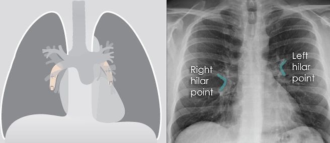
2. Identify the hilar point: Look for the site where the most superior upper lobe vessel – either vein or artery – crosses the lateral margin of the little finger. The point of crossing is known as hilar point and forms a horizontal “vee” (> or <).
Interpretation: The left hilum must never be lower than the right hilum. Whenever a left hilum appears lower than the right hilum – look for other evidence suggestive of:
- Collapse of either the left lower lobe or of the right upper lobe
- Enlargement of the right hilum
b. Analyze the density of hilum:
The presence of more than a slight difference in density raises the suspicion that there is abnormal tissue at the hilum — e.g. a hilar mass.
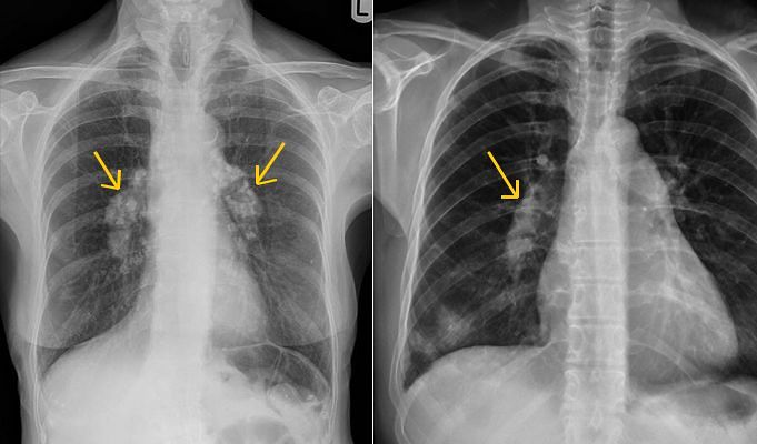
c. Analyze the enlargement of hilum (if present):
1. Lymph Node enlargement:
- Lobulated appearance
- Presence of calcification within the mass indicates lymph node enlargement usually tuberculosis. Egg shape calcification indicates lymphadenopathy due to silicosis.
2. Arterial enlargement:
- Can be traced into and are seen to be part of the “mass” – hilar mass
- Smooth margins
- In pulmonary arterial hypertension the arteries in the outer two-thirds of each lung are disproportionately smaller (diameter) than those at the hila (peripheral pruning)
Right: Pulmonary artery hypertension
3. Malignancy:
- Spiculated irregular or indistinct margins
- Hilar enlargement due to malignant lung lesion is also associated with superior mediastinal lymphadenopathy. Look at the lung fields (for presence of tumor) and bone/ribs for metastasis.
Causes of Hilar Enlargement:
Unilateral:
- Infection: tuberculosis, viral infection in children
- Vascular: pulmonary artery stenosis, pulmonary artery aneurysm
- Tumour: lymph nodes (metastases; lymphoma; bronchial carcinoma)
Bilateral:
- Sarcoidosis
- Tumour: metastases, lymphoma
- Vascular: pulmonary arterial hypertension (COPD; mitral valve disease; left to right shunt; recurrent pulmonary embolism)
- Infection: tuberculosis (occasionally)
Hilum convergence/bifurcation sign: Helps to analyze if the hilar enlargement is due to –
- Mass: If the vessels converge medial to the hilar shadow
- Vessels: If the vessels converge and merge directly on to the hilar shadow
Other signs of hilum:
Silhouette sign: This role of this sign is not just limited for the diagnosis of consolidation but to any mass lesion projected over a hilum.
- Hilar mass: Obscure the adjacent margin of the arteries at the hilum
- Mass – anterior or posterior to hilum: Margins stay unobscured
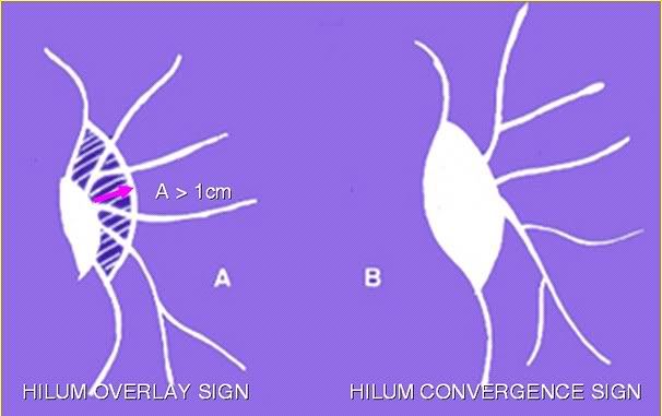
Hilum overlay sign: Cardiac enlargement vs Mediastinal enlargement
- Hilum pushed lateral to the lateral border of the “mass” — cardiac enlargement.
- Hilum smedial to the lateral border of the “mass” — mediastinal mass present.