
Vestibule and Sensory receptors
Location: Medial to tympanic membrane and Posterior to Cochlea
Sensory receptors
1. Macula:
- Present in otolith (calcium carbonate crystals) organs – saccule (anteriorly) and utricle (posteriorly)
- Both are connected by corresponding ducts, which together will form endolymphatic duct, this passes through a bony canal (the vestibular aqueduct), and expands into flattened endolymphatic sac, blending into posterior cranial fossa dura.
- Picks up linear acceleration:
- Utricle have Upright orientation of hair cells and hence, the cilia is displaced in horizontal plane (left-right, front-back)
- Saccule have Slanted orientation hair cells and hence, the cilia is displaced in vertical plane (up-down)
2. Crista:
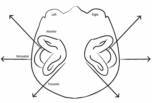
- Present in semicircular canals
- 3 semicircular canals – 2 vertically and 1 horizontally arranged
- Anterior/Superior semicircular canal (vertically; anterolateral)
- Posterior semicircular canal (vertically, posterolateral)
- Horizontal semicircular canal (horizontally)
- Corresponding semicircular canals in right and left are at 90° to eachother.
- Each semi-circular canal has 2 ends:
- Dilated ampullary end (contains crista)
- Non-dilated non-ampullary end (joined to anterior and posterior semicircular canal to form crus-commune)
- Picks up circular acceleration (rotatory motion)
Remember: “C” for crista, “C” for canals and “C” for circular motion
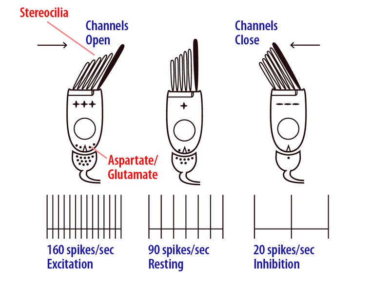
Both macula and crista have sensory hair cells graded in height:
- Tallest: Kinocilium
- Others: Sterocilia
Firing:
- Deflection of sterocilia towards kinocilium: depolarization
- Deflection of sterocilia away from kinociluim: hyperpolarization
Stimulation of semicircular canal as per head motion:
- Horizontal semicircular canal: Kinocilium is lateral to sterocilia
- Left head-turn depolarizes left horizontal semicircular canal and hyperpolarizes right horizontal semicircular canal
- Right head-turn depolarizes right horizontal semicircular canal and hyperpolarizes left horizontal semicircular canal
- In Anterior semicircular canal – Kinocilium is anterior to sterocilia and In Posterior semicircular canal – Kinocilium is posterior to sterocilia
- Forward head tilt (without left or right) depolarizes anterior semicircular canal and hyperpolarizes posterior semicircular canal
- Forward-left head tilt depolarizes left anterior semi-circular canal and right posterior semicricular canal and hyperpolarizes left posterior semi-circular canal and right anterior semi-circular canal and vice-versa
Note:
- Semi-circular canal towards the direction of head turn is activated (right or left and anterior or posterior).
- Activation of one semicircular canal in a side, deactivates corresponding semicircular canal in other side.
- Activation of anterior semicircular canal in a side, deactivates posterior semicircular canal in the same side and vice-versa.
- Pairing A/P semicricular canals: Left anterior and Right posterior; Right anterior and Left posterior
Vestibular Nerve
The nerves from the semicircular canals and the otolithic organs project to the Scarpa’s Ganglion in Internal acoustic meatus.
Axons of the Scarpa’s ganglion form the vestibular nerve (CN VIII).
Vestibular Nuclei
Location: Ponto-medullary junction
4 divisions of vestibular complex:
- Superior vestibular nuclei of Bechterew
- Medial vestibular nuclei of Schwalbe (Least specialized) – sends afferent and efferent to all the pathways
- Lateral vestibular nuclei of Dieter
- Inferior vestibular nuclei
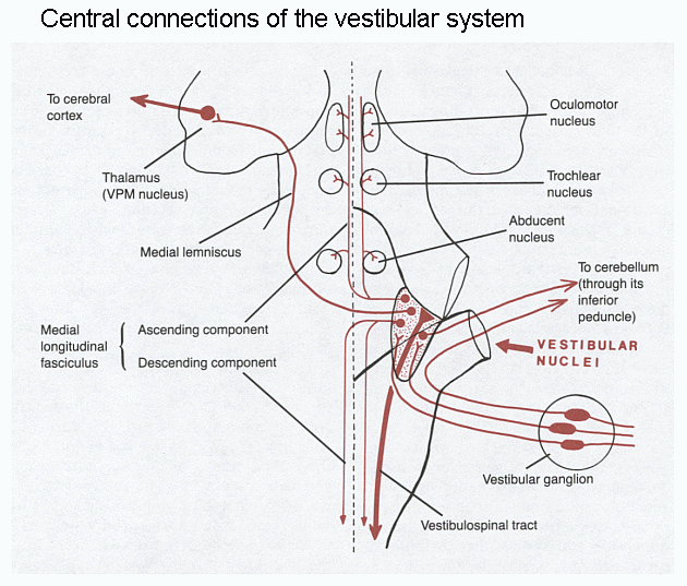
Vestibular fibers
Medial vestibular nuclei is involved in all the pathways; besides, we will discuss only the major pathway of efferents from the specific vestibular nucleus:
1. Superior vestibular nucleus: Vestibulo-occular reflex pathway (Stabilize eye gaze)
2. Medial vestibular nucleus: Medial vestibulo-spinal pathway (Stabilize posture)
3. Lateral vestibular nucleus: Lateral vestibulo-spinal pathway (Stabilize posture)
4. Inferior cerebellum nucleus: Vestibulo-cerebellar pathway (Cerebellum)
Vestibulo-Ocular Reflex (VOR)
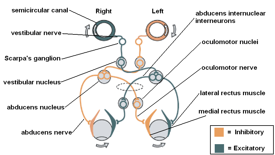
- Fibers activate CONTRALATERAL CN VI nucleus and inhibiti IPSILATERAL CN VI nucleus
- CN VI will activate CONTRALATERAL CN III (i.e. ipsilateral to vestibular system stimulated) through Medial Longitudinal Fasciculus (MLF)
Function: stabilizes images on the retina during head movement by producing an eye movement in the direction opposite to head movement, thus preserving the image on the center of the visual field.
Example:
Left head turn → Left vestibular nuclei and nerve stimulated → Right CN VI nuclei and lateral rectus stimulated → Left CN III and medial rectus stimulated → Right lateral rectus turns right eye to right and Left medial rectus turns left eye also to the right i.e. when head is turned to left, both the eyes fix gaze to right.
Nystagmus in Unilateral Vestibular Damage:
Unopposed action of Contralateral Vestibular nerve and nuclei leads to:
- Vestibulo-ocular reflex (Slow component): Eyes slowly look to side of vestibular damage
- Attempt of cerebral correction (Fast component): Eyes rapidly move back to side away from vestibular damage
Caloric testing:
Lateral or horizontal semicircular canal is oriented vertical by elevating the head 30° from horizontal.
- Warm water → Stimulates horizontal semicircular canal on same side and inhibits the same on opposite side → Eye moves to opposite side by vestibulo-ocular reflex (slow component) → Correction by moving eye back to same side (fast component) i.e. Nystagmus to the same side
- Cold water → Inhibits horizontal semicircular canal on same side and stimulates the same on opposite side → Eye moves to the same side by vestibuo-ocular reflex (slow component) → Correction by moving eye to the oppoiste side (fast component) i.e. Nystagmus to the opposite side
Mnemonic: COWS (Cold Opposite and Ward Same side)
Vestibulo-Spinal Pathway
Medial vestibulospinal pathway:
Efferent medial vestibular nucleus fibers → descend in MLF → become medial vestibulo-spinal tract → cervical and upper thoracic motor nuclei
Function: Stabilize Head and neck posture
Lateral vestibulospinal pathway:
Efferent lateral vestibular nucleus fibers → descend on anterior horn of spinal cord as lateral vestibulospinal tract
Function: Forelimb antigravity posture
Vestibulo-cerebellar Pathway
Involved part of cerebellum: Mildine cerebellum (Archicerebellum)
Direct vestibulocerebellar tract:
- Vestibular labyrinth → fibers directly to vermis in the midline cerebellum
Indirect vestibulocerebellar tract:
- Vestibular labyrinth → Inferior vestibular nucleus → ipsilateral inferior cerebellar peduncle → uvula and flocculonodular lobe in midline cerebellum
Midline cerebellum → Efferent fibers → Bilateral vestibular nucleus complex
Vestibulo-cerebral Pathway
Efferent vestibular projections to bilateral Ventral Posterior group of thalamus
Cortical regions of the brain known to be involved with vestibular processing:
- Frontal eye fields: control eye movements and receive vestibular motion information
- Primary somatosensory cortex (Areas 2v and 3a): map body location and movement signals
- PIVC (Parieto-Insular Vestibular Cortex): responds to body and head motion information
- Posterior parietal cortex: motion perception and responds to both visual and vestibular motion cues
- Hippocampus and parahippocampul regions: spatial orientation and navigation functions
Vestibulo-autonomic Pathway
- Some vestibular efferent projections to reticular formation, dorsal pontine nuclei, and nucleus of solitary tract.
- Function: Stabilize respiration and blood pressure during body motion and changes relative to gravity
- Role in motion sickness