

| Site | Point of insertion | Direction | Indications |
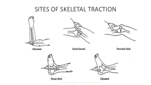
| Olecranon (K-wire) | 1.25 inches (3 cm) distal to olecranon tip – deep to subcutaneous border of upper ulna (avoids ulnar joint and open epiphysis) | Medial to Lateral – At right angles to longitudinal axis of ulna (Avoids ulnar nerve) | Supracondylar or Distal humerus fractures |
| 2nd and 3rd metacarpals (K-wire) | 1 inch (2-2.5 cm) proximal to distal end of 2nd metacarpal | Radial to ulnar, traversing 2nd and 3rd metacarpal diaphysis – At right angles to the longitudinal axis of radius | Difficult reduction forearm or distal radius fracture |
| Greater trochanter (Steinmann or Denham’s pin) | 1 inch (2.5 cm) below the most prominent part of greater trochanter – midway between anterior and posterior surface of femur | Lateral to Medial | Central fracture dislocation of hip |
| Distal femur (Steinmann or Denham’s pin) | 3 cm proximal to lateral joint line (i.e. just proximal to femoral condyle or at the level of proximal pole of patella in relaxed and extended knee) – avoids lateral knee joint capsule which reaches 1.25-2cm above knee joint, distal femoral physis | Lateral to Medial – pin should pass along or slightly posterior to the midcoronal plane of femoral shaft and pass just proximal to the adductor tubercle in order to avoid engagement of the collateral ligaments (Traditionally medial to lateral direction suggested to save femoral artery in Hunter’s canal) | Superior force acetabular fractures and femoral shaft fractures |
| Proximal tibia -Perkin’s traction (Steinmann or Denham’s pin) | 2cm behind and 2cm below tibial tuberosity (to avoid proximal weaker cancellous bone and to avoid common peroneal nerve distally as it courses anteriorly after winding around fibular nerve) | Lateral to Medial (to avoid common peroneal nerve) | Fractures of tibia and femur from subtrochanteric region distally |
| Distal tibia | 5cm above the ankle joint – midway between anterior and posterior borders of tibia | Medial to Lateral (avoid saphenous vein) | Tibial plateau fracture |
| Calcaneus | 1.5 inches (4 cm) inferior and posterior to medial malleolus (avoid tendons, neurovascular bundle passing behind the malleoli and subtalar joint) | Medial to Lateral | Tibial shaft fracture or calcaneal fracture |