
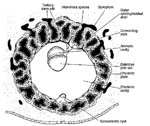
In the Week 2, we had a bilaminar germ disc with 2 cavities (amniotic cavity and yolk sac) suspended in the chorionic (extraembryonic cavity) by a connecting stalk (future umbilical cord).
In the 3rd week, the embryo will have 3 germ layers (ectoderm, mesoderm and endoderm) following a process called gastrulation.
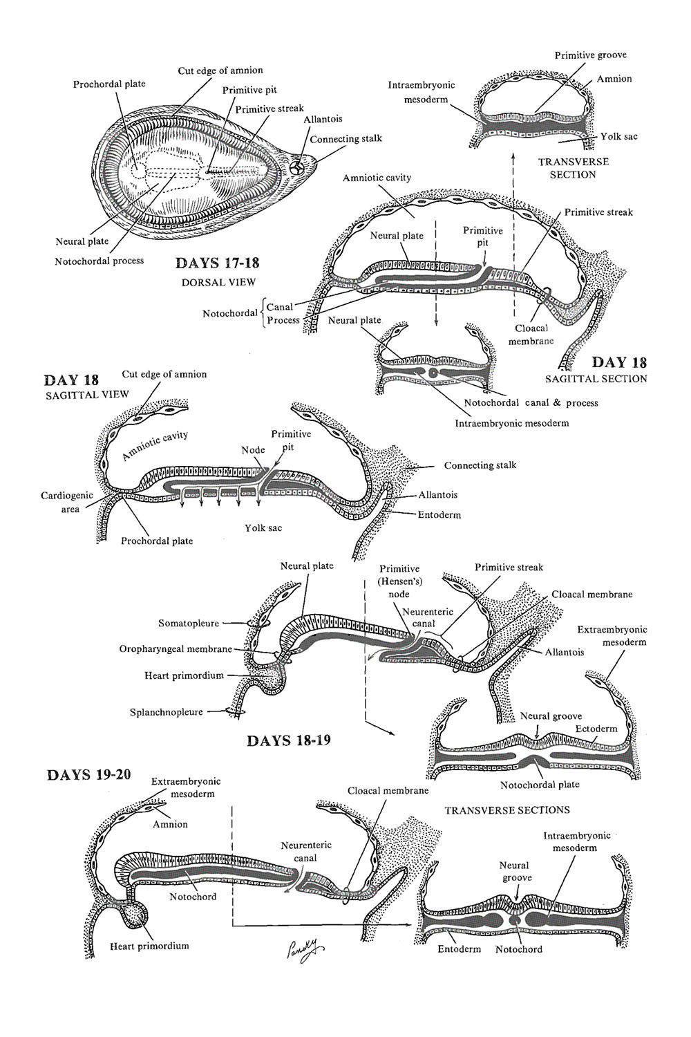
Primitive Streak
- A linear band of thickened epiblast that grows caudo-cranially (towards prechordal plate)
- Arises due to proliferation and migration of epiblasts in the direction of median line
- Consists of: primitive groove and a primitive pit contained in a primitive node cranially
Invagination of Epiblast cells
Cells of the epiblast migrate toward primitive streak and detach from epiblast layer:
- Some displace hypoblast: Embryonic endoderm
- Others come to lie between epiblast and newly created endoderm: Embryonic mesoderm
- Remaining epiblast layer: Embryonic ectoderm
Note: Mesoderm lies between the ectoderm and endoderm as a continuous sheet except at the buccopharyngeal membrane and cloacal membrane. These membranes have ectoderm and endoderm only and will lie at the rostral (head) and caudal (tail) of the gastrointestinal tract.
- Buccopharyngeal membrane: future mouth
- Cloacal membrane: future anus and genital openings
- Cardiogenic region (cranial to buccopharyngeal membrane): future heart and ventral chest
- Septum transversum (most cranial part of the disc): contribute to diaphragm and separates the heart from liver
Embryonic and Extraembryonic mesoderm meet: This occurs by the overflow of invaginating mesodermal cells that migrate slowly out of the margins of the embryonic disc to come in contact with the extraembryonic mesoderm covering the yolk sac and amniotic cavity.
Prechordal plate forms: Early epiblast cells invaginating through the primitive node and migrating in cephalic direction forms prechordal plate between primitive node and buccopharyngeal membrane.

Notochord Formation
Formation of notochordal process (Day 16-18): Epiblast cells continues to migrate cranially through the primitive node towards prechordal pate to form notochordal process.
Hence, notochord is a mesoderm located between prechordal plate and primitive node that gives rise to nucleus pulposus in future.
Formation of neurenteric canal and notochordal plate:
- Canalization of notochordal process by extension of primitive pit cranially
- The floor of canalized notochord and underlying hypoblast degenerate
- Formation of neurenteric canal between notochordal canal and yolk sac establishing a communication between them.
- The remaining roof of notochordal process remains intercalated in the hypoblast and is called notochordal plate.
Formation of definitive notochord:
The epiblast cells migrating to become endoderm replaces the hypoblast as discussed earlier. During this process, the notochordal plate also grows and separates from the endoderm to form definitive notochord.
Extent: Primitive pit to Prechordal plate
Stimulation of neurulation by notochord and paraxial mesoderm:
- Around day 18, the ectoderm over the developing notochord thickens to form Neural plate.
- Neural plate extends cranially along with the neural plate.
- By the end of 3rd week: lateral edges of neural pate forms a groove (neural groove) by elevating a fold (neural fold)
- The closure of the neural fold begins and ends during the 4th week of development.
Development of Intraembryonic circulatory system
Prepares for the development of heart that occurs in 4th week –
Primary villi: Growth of cytotrophoblast into the syncytiotrophoblast forming villous structure.
Secondary villi: Primary villi obtain a mesenchymal core
Tertiary villi or Definitive placental villus: Small capillaries arise in the mesenchymal core in secondary villi
Intraembryoninc circulatory system: Villous capillaries make contact with capillaries developing in chorionic plate and connecting stalk.
Allantois
Around day 16, allantois appears as a diverticulum from the caudal wall of the yolk sac. It is involved in early blood formation
Germ Layer Derivatives
We have discussed earlier, the rule mnemonic to remember the germ layer derivatives.
Establishment of Body Axes
Occurs: before and during the period of gastrulation.
Antero-posterior axis:
Craniocaudal patterning of the embryonic axis is controlled by homeobox genes.
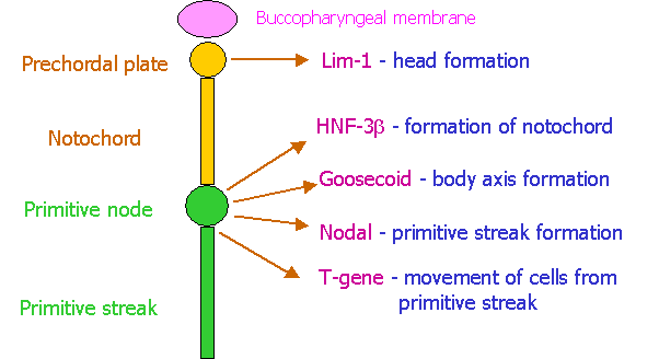
Overexpression of goosecoid gene hase been linked to conjoined twinning.
Right-left axis:
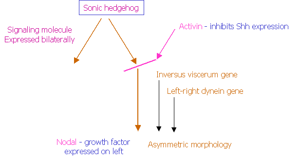
Left side/right side (L/R) axis determination begins with the asymmetric activity sonic hedgehog protein (Shh) only on the future left side since, Shh activity is suppressed on the future right side by activin.
In addition, the neurotransmitter serotonin (5HT) plays an important role in L/R axis determination. 5-HT is concentrated on left site because it is broken down by MAO on right side. There are also genes that specifically determine development of structures only on the left side – Inversus viscerum and left-right dynein. Mutations of these genes cause “situs inversus”.
Clinical Correlate
Chordoma
- Benign or malignant tumor arising from the remnants of notochord.
First Missed Menstrual Period
- Week 3 of embryonic development coincides with this period.
Selective Serotonin Reuptake Inhibitors (SSRIs)
- Teratogenic – increased risk of heart malformations due to role of serotonin in establishing laterality.
Sacrococcygeal teratoma
- Tumor that arises from the remnants (pluripotent cells) of primitive streak (which normally starts degenerating by 4th week of development).
Caudal dysplasia (Sirenomelia)
- Results from abnormal gastrulation – migration of mesoderm in caudal-most region of embryo is disturbed. Because, mesoderm contributes to formation of limbs, urogenital system and lumbosacral vertebrae, abnormalities in these structures ensue. It is associated with VATER or VACTERL syndrome.