Skip to content
Epomedicine

Mnemonics, Simplified Concepts & Thoughts

Epomedicine

Mnemonics, Simplified Concepts & Thoughts

Ligaments of Pelvis

Epomedicine, Oct 3, 2020Oct 3, 2020

Inherent stability of the pelvis is provided by ligaments. The 3 groups of ligaments are:

1. Sacrum to Pelvis:

Sacroiliac ligamentous complex: is divided into posterior (short and long) and anterior ligaments. Posterior ligaments provide most of the stability.

Sacrotuberous ligament: runs from the posterolateral aspect of the sacrum and the dorsal aspect of the posterior iliac spine to the ischial tuberosity.

Sacrospinous ligament: is triangular, running from the lateral margins of the sacrum and coccyx and inserting on the ischial spine.

pelvic ligaments

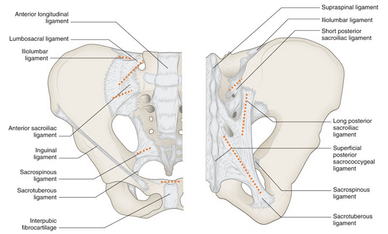
2. Pubis to pubis: Symphyseal ligaments

3. Lumbar spine to pelvic ring: Provides additional stability

Iliolumbar ligaments: originate from the L4 and L5 transverse processes and insert on the posterior iliac crest.

Lumbosacral ligaments: originate from the transverse process of L5 to the ala of the sacrum.

Transversely placed ligaments: resist rotational forces

  1. Anterior sacroiliac ligament
  2. Short posterior sacroiliac ligament
  3. Iliolumbar ligament
  4. Sacrospinous ligament

Vertically placed ligaments: resist vertical shear forces

  1. Long posterior sacroiliac ligament
  2. Sacrotuberous ligament
  3. Lateral lumbosacral ligament

Injured ligaments of the pelvis determine relative contributions to pelvic stability:

  • Symphysis alone: pubic diastasis <2.5 cm
  • Symphysis and sacrospinous ligaments: >2.5 cm of pubic diastasis (rotationally unstable)
  • Symphysis, sacrospinous, sacrotuberous, and posterior sacroiliac: unstable vertically, posteriorly, and rotationally
32 shares
  • Facebook32
  • Twitter
PGMEE, MRCS, USMLE, MBBS, MD/MS AnatomyMusculoskeletal systemOrthopedics

Post navigation

Previous post
Next post

Related Posts

PGMEE, MRCS, USMLE, MBBS, MD/MS

Hilar Arrangement in Lungs, Liver, Kidneys : Mnemonic

Jun 4, 2023Jun 4, 2023

Hilum is an indentation in the surface of a kidney, spleen, or other organ, where blood vessels, ducts, nerve fibers, etc. enter and leave it. Hilum of Lung Mnemonic: VAD (from anterior to posterior) Hilum of Liver (Porta Hepatis) Mnemonic: DAVE (from anterior to posterior) Hilum of Kidney Mnemonic: VAD…

Read More
PGMEE, MRCS, USMLE, MBBS, MD/MS

Spinal segments vs Vertebral level

Aug 9, 2023Aug 9, 2023

Spinal segment: The part of spinal cord to which a pair of spinal nerves (right and left) is attached. Cause of mismatch between spinal segments and vertebral level: General rule: Vertebral level Add Spinal segment Cervical + 0 Same C7-T1 C8 T1-T3 +1 T1-T4 T3-T6 +2 T5-T8 T7-T8 +3 T9-T11…

Read More
PGMEE, MRCS, USMLE, MBBS, MD/MS macrophage

Tissue Specific and Named Macrophages

Jan 17, 2017Jan 19, 2017

Subtle differences in the morphology and functions of macrophages develop as a result of the influence of a particular microenvironment. Appearance of macrophages to histologists have been described as a kind of mythological Proteus, “a creature who had the power of changing his appearance at will”. The life-span of these fixed…

Read More

Leave a Reply Cancel reply

Your email address will not be published. Required fields are marked *

This site uses Akismet to reduce spam. Learn how your comment data is processed.

Pre-clinical (Basic Sciences)

Anatomy

Biochemistry

Community medicine (PSM)

Embryology

Microbiology

Pathology

Pharmacology

Physiology

Clinical Sciences

Anesthesia

Dermatology

Emergency medicine

Forensic

Internal medicine

Gynecology & Obstetrics

Oncology

Ophthalmology

Orthopedics

Otorhinolaryngology (ENT)

Pediatrics

Psychiatry

Radiology

Surgery

RSS Ask Epomedicine

  • What to study for Clinical examination in Orthopedics?
  • What is the mechanism of AVNRT?

Epomedicine weekly

  • About Epomedicine
  • Contact Us
  • Author Guidelines
  • Submit Article
  • Editorial Board
  • USMLE
  • MRCS
  • Thesis
©2026 Epomedicine | WordPress Theme by SuperbThemes