Skip to content
Epomedicine

Mnemonics, Simplified Concepts & Thoughts

Epomedicine

Mnemonics, Simplified Concepts & Thoughts

pleural effusion xray

Chest X-ray – Pleural Effusion

Epomedicine, Jun 21, 2015

Pleura is a mesothelial lined sac that envelopes the lungs and comprises of 2 membranous walls i.e. visceral pleura and parietal pleura that encloses pleural space filled with pleural fluid. Pleural space contains about 0.3 ml/kg body weight of pleural fluid. The pleura is not visible on a normal CXR except where it forms part of a lung fissure or where the two lungs abut each other in the midline
pleural effusion xray

Erect frontal Chest X-ray:

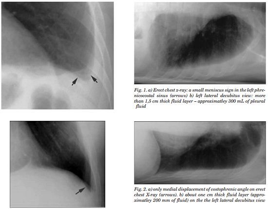
  1. Obliteration of costophrenic angle: requires 200-300 ml pleural fluid
    • Concave meniscus (horizontal in case of hydropneumothorax)
  2. Massive pleural effusion: opacification of entire hemithorax and shifting of mediastinum to the opposite side
    • Causes “white-out” lung
    • Around 5-7 l of pleural fluid
  3. Lamellar pleural effusion: linear opacification paralleling the lateral aspect of lung
  4. Encysted pleural effusion: loculation within a fissure or elsewhere
    • Can be mistaken for lung tumor
  5. Subpulmonic effusion:
    • left: widening of the distance between gastric bubble and the superior margin of hemidiaphragm (normally < 7mm)
    • right: peak of the hemidiaphragm is shifted laterally

Lateral Chest X-ray:

Lateral films are able to identify a smaller amount of fluid as the costophrenic angles are deepest posteriorly

  • Can detect an effusion as small as 50 ml

Supine Chest X-ray:

Due to the effect of gravity, the pleural fluid is distributed throughout the posterior part of the pleural during supine position – this cause the hemithorax to appear whiter or paler grey compared to the normal side. Vessels are often visible through the shadowing.

  • Requires about 200 ml fluid
23 shares
  • Facebook23
  • Twitter
PGMEE, MRCS, USMLE, MBBS, MD/MS Internal medicinePediatricsRadiologyRespiratory system

Post navigation

Previous post
Next post

Related Posts

PGMEE, MRCS, USMLE, MBBS, MD/MS jugular foramen

Structures passing through Jugular foramen : Mnemonic

Jun 29, 2018

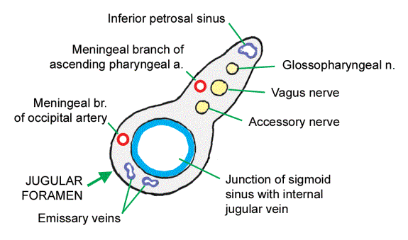
Mnemonic: IPS 9,10,11 MAP JOE Anterior part: IPS Inferior Petrosal Sinus Middle part: 9, 10, 11 MAP CN IX CN X CN XI Meningeal br. of Ascending Pharyngeal artery Posterior part: JOE Jugular vein (internal; lower end of sigmoid sinus) Occipital artery Emissary vein Vernet’s syndrome (Jugular foramen syndrome) Unilateral…

Read More
PGMEE, MRCS, USMLE, MBBS, MD/MS

Column Concept of Acetabulum

May 26, 2020Jun 3, 2020

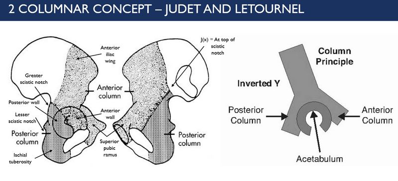
2 Column Concept or Principle of Acetabulum (Letournel and Judet) According to this concept, “C” shaped acetabular structure is contained between the limbs of lamda [λ] or inverted “y”. The longer limb represents anterior column and the shorter limb represents the posterior column. The anterior and posterior walls are extensions…

Read More
PGMEE, MRCS, USMLE, MBBS, MD/MS

AKI RIFLE Criteria Mnemonic

Aug 6, 2024Aug 6, 2024

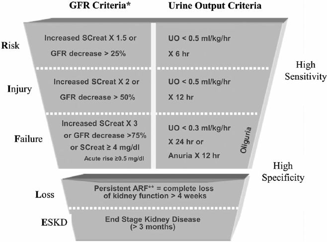
Stage GFR criteria Urine output criteria Mnemonic 1, 2, 3, 4 1, 2, 3 1. Risk Cr ↑ 1.5-2 XGFR ↓ 25-50% (>1/4) <0.5 ml/kg/hr X 6-12 (>1/4 day) 2. Injury Cr ↑ 2-3 XGFR ↓ >50-75% (>2/4) <0.5 ml/kg/hr X >12-24 hrs (>2/4 day) 3. Failure Cr ↑ >3…

Read More

Leave a Reply Cancel reply

Your email address will not be published. Required fields are marked *

This site uses Akismet to reduce spam. Learn how your comment data is processed.

Epomedicine. Chest X-ray – Pleural Effusion [Internet]. Epomedicine; 2015 Jun 21 [cited 2026 Sep 2]. Available from: https://epomedicine.com/medical-students/chest-x-ray-pleural-effusion/.

Pre-clinical (Basic Sciences)

Anatomy

Biochemistry

Community medicine (PSM)

Embryology

Microbiology

Pathology

Pharmacology

Physiology

Clinical Sciences

Anesthesia

Dermatology

Emergency medicine

Forensic

Internal medicine

Gynecology & Obstetrics

Oncology

Ophthalmology

Orthopedics

Otorhinolaryngology (ENT)

Pediatrics

Psychiatry

Radiology

Surgery

RSS feed: Ask Epomedicine Ask Epomedicine

  • What to study for Clinical examination in Orthopedics?
  • What is the mechanism of AVNRT?

Epomedicine weekly

  • About Epomedicine
  • Contact Us
  • Author Guidelines
  • Submit Article
  • Editorial Board
  • USMLE
  • MRCS
  • Thesis
©2026 Epomedicine | WordPress Theme by SuperbThemes