Table of Contents
Follow the general principles of orthopedic examination outlined in the article below.
Standing
1. Note general point of pain: C sign (Patients with intra-articular hip pathology often localize the area of pain by cupping the thumb and index fnger in the shape of the letter C above the affected area.)
2. Gait (Watch 6-8 stride lengths): Pelvic tilt/rotation, Stride length, Stance phase, FPA
3. Single leg stance phase test: Neural loop/Abductor strength
4. Inspection:
- Body habitus
- Spinal alignment: Shoulder-iliac crest heights, lordosis, scoliosis (towards affected side in abduction deformity, towards normal side in adduction deformity; hint: side wherever ASIS is lower) , leg length (tilted pelvis, equinus on shorter side, knee flexion on longer side)
- Symmetry of gluteal folds: Asymmetrical on developmental dysplasia of hip, muscular dystrophy or leg length discrepancy
- Forward bending (Structural vs Nonstructural scoliosis; ROM) and lateral bending ROM
- Laxity: Beighton scale
Seated
5. Vascular and Lymphatic examination: Dorsalis pedis and posterior tibial artery pulse; Skin for swelling, scarring or side-to-side asymmetry
6. Neurologic: Sensory and Motor function and DTR (Patellar, Achilles)
7. Passive straight leg raise: Radicular neurologic symptoms
8. Passive hip rotation: Seated position ensures that the ischium is square to the table, thus providing adequate stability at 90 degrees of hip flexion
- Internal rotation: Normal 20-35 degrees
- External rotation: Normal 30-45 degrees
Supine
9. Inspect for leg length discrepancies (LLD)
10. Palpate:
- Bony landmarks: ASIS, Iliac crest, Greater trochanter, Symphysis pubis, Pubic tubercle, Adductor tubercle
- Local temperature
- Joint tenderness:
- Anteriorly: 2 cm below and lateral to mid-inguinal point
- Posteriorly: Junction of lateral 1/3 and medial 2/3 of a line joining GT and PSIS
- Vascular sign of Narath (Femoral pulse)
- Inguinal lymph nodes
- Abdomen
11. Range of Motion (ROM): Flexion, abduction, adduction
| Range of motion assessment | Normal (degrees) | Center | Stationary arm | Moving arm |
| Seated internal rotation | 20-35 | Center of patella | Aligned vertically | Line to tibial crest and midway between malleoli |
| Seated external rotation | 30-45 | Center of patella | Aligned vertically | Line to tibial crest and midway between malleoli |
| Extended internal rotation | 20-35 | Center of patella | Aligned horizontally | Line to tibial crest and midway between malleoli |
| Extended external rotation | 30-45 | Center of patella | Aligned horizontally | Line to tibial crest and midway between malleoli |
| Supine hip flexion | 100-110 | GT | Lateral midline of pelvis | Line to lateral femoral epicondyle |
| Adduction | 20-30 | ASIS | Line to other ASIS | Line to midline patella |
| Abduction | 45 | <45 | Line to other ASIS | Line to midline patella |
12. Fixed adduction and abduction deformity:
- ASIS at both sides at same level: Pelvis square
- ASIS at affected side higher: Fixed adduction deformity
- ASIS at affected side lower: Fixed abduction deformity
- Squaring of pelvis: Bring the ASIS into same level by further adducting in adduction deformity and abducting in the abduction deformity
- Quantifying the fixed adduction and abduction deformity:
- Squaring the pelvis: Angle of adduction or abduction with the vertical line
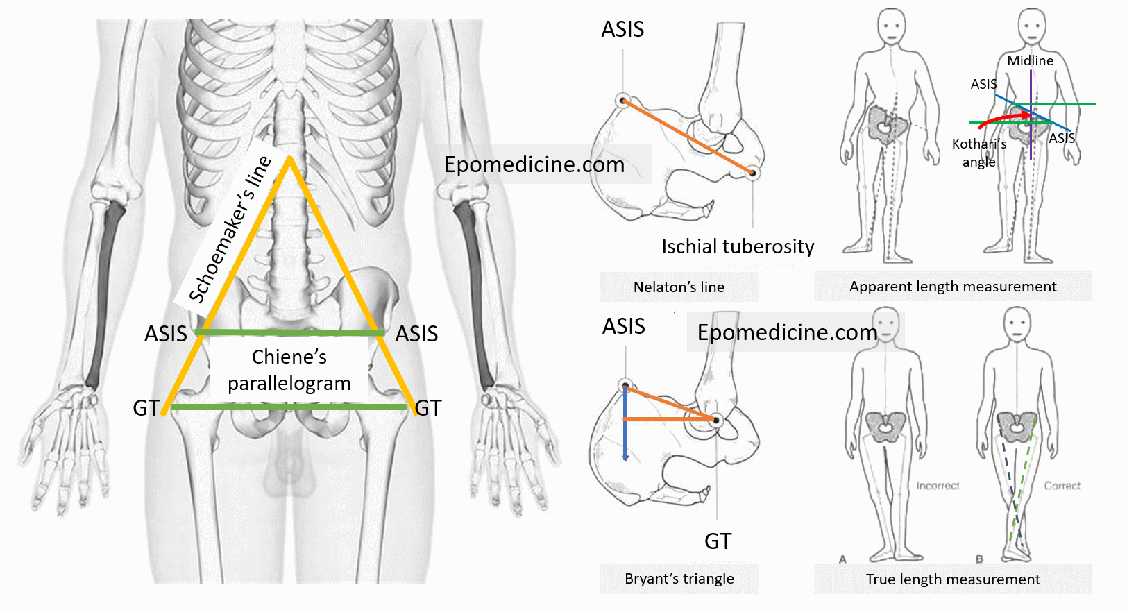
- Kothari’s angle: A line is drawn from suprasternal notch to symphysis pubis. Perpendicular is dropped from ASIS to this line and the angle between this line and the line connecting the ASIS on two sides form the Kothari’s angle.
13. Thomas test
14. FADDIR:
- Hip pain: Anterior femoroacetabular impingement, Torn labrum
- Pain along the piriformis distribution or sciatic nerve pain down the leg: Piriformis syndrome
15. FABER (Patrick test; Figure of 4 position): Distinguish between back and hip pathology, specifically sacroiliac joint
- Sacoiliac discomfort: Sacroiliitis
- Recreation of hip pain: Posterior femoroacetabular impingement, ligamentous injury or trochanteric pathologies
16. DIRI test and DEXRIT
| DIRI test | DEXRIT |
| Dynamic internal rotation impingement test | Dynamic external rotation impingement test |
| Establish 0 pelvic set point and eliminate lumbar lordosis by holding nonaffected leg in flexion beyond 90 degrees – examined hip is brought into 90 degrees of flexion or beyond and is passively taken through a wide arc of adduction and internal rotation (dynamic FADDIR) | Establish 0 pelvic set point and eliminate lumbar lordosis by holding nonaffected leg in flexion beyond 90 degrees – examined hip is brought into 90 degrees of flexion or beyond and is passively taken through a wide arc of abduction and external rotation (dynamic FABER) |
| Positive – Recreation of hip pain | Positive – Recreation of hip pain |
| Anterior FAI, torn labrum | Superior FAI, torn labrum |
17. Posterior rim impingement test (EABER): Posterior FAI, torn labrum
The patient is positioned at the edge or end of the examination table so that the leg can hang freely to full extension. The patient established the pelvic zero set-point with both legs held in flexion. The examined leg is allowed to reach full extension off the table, and then taken into abduction and external rotation.
18. Passive supine rotation (log roll) test
Gently roll the limb into internal rotation and external rotation. Pain suggests intra-articular hip pathology.
Dial test: Passive internal rotation of the leg, followed by releasing the leg and allowing it to external rotate. External rotation of more than contralateral limb or more than 45°, relative to vertical, is a positive test and indicates iliofemoral ligament laxity.
19. Heel strike: Trauma or femoral fracture
20. Straight leg raise against resistance (to 45 degrees; Stinchfield test): Pain (intra-articular pathology as the iliopsoas presses against the acetabular labrum during active resistance) or Iliopsoas weakness
21. Length measurements:
- Apparent length measurement: Place the legs parallel; Midline point of body (xiphisternum or umbilicus) to tip of medial malleolus (adduction makes the limb appear shorter – each 10 degrees of fixed adduction/abduction add a further 3 cm change in apparent length to any real shortening)
- True length measurement: Place the normal limb in a comparable position of adduction or abduction to the abnormal limb; ASIS to medial malleolus (if there is a fixed deformity, the good leg must be placed in a comparable deformed position relative to the pelvis)
- Is the shortening above or below the knee
- Segmental measurements: Measure –
- Femur: from ASIS to medial knee joint line
- Tibia: from medial knee joint line to tip of medial malleolus
- Galeazzi’s test: If both knees are flexed while the heels remain together on the couch, it can be seen whether the shortening is in the femur or tibia.
- Knee lies lower down: Tibial shortening
- Knee lies proximally: Femoral shortening
- Segmental measurements: Measure –
- If the shortening is in femur, is it above the trochanter or below the trochanter?
- Bryant’s triangle: Not helpful in bilateral hip joint pathology
- Chiene’s parallelogram: 2 lines joining the GT on both sides and ASIS on both sides must be parallel (supratrochanteric shortening at 1 side leads to convergence of line)
- Schoemaker’s line: The lines passing through GT to ASIS on both sides normally meets in the midline above the umbilicus
- Unilateral supratrochanteric shortening: Meet away from the midline on opposite side
- Bilateral supratrochanteric shortening: Meet in midline but inferior to umbilicus
a. True shortening = Apparent shortening: No compensation
b. True shortening > Apparent shortening: Partial compensation with Fixed abduction deformity
c. True shortening < Apparent shortening: Fixed adduction deformity without compensation
22. Telescopic test: Stabilize the pelvis by pressing on ASIS with thumb and thenar eminence of one hand while the fingertips touch the greater trochanter. Flex the affected hip and knee to 90 degrees and grasp the distal thigh and knee between your chest and right armpit. Push and pull along the longitudinal axis of femur. Note the amount of excursion of the tip of trochanter with fingertips and compare to the opposite side. It may be positive in:
- Nonunion neck of femur with resorbed fracture ends
- Old unreduced posterior dislocation of hip
- DDH
- Pathological dislocation of hip
- Girdlestone arthroplasty
- Septic sequelae of hip
Lateral
23. Palpation:
- Suprasacroiliac area, SI joint, gluteus maximus origin, piriformis muscle
- Facets of greater trochanter (gluteus minimus inserts at anterior facet, gluteus medius inserts at posterosuperior and lateral facet, trochanteric bursa at posterior facet)
- Sciatic nerve (Sciatic point): midway between ischial tuberosity and posterior border of trochanter with hip flexed (moves gluteus maximus away)
- Tenderness: Herniated lumbar disc, Spasm of piriformis, Direct trauma over nerve
24. Abductor strength:
- Overall: With knee in extension
- Gluteus medius: With knee in flexion (to release gluteus maximus contribution for the iliotibial band)
- Gluteus maximus: Abducting and extending the hip
25. Ober’s test
26. Ischiofemoral impingement test (HEADER; Hip extension, abduction, external rotation)
27. Nelaton’s line: A measuring tape of string is stretched from ischial tuberosity to ASIS with hip in 90 degrees flexion. Normally, greater trochanter lies on or below the line. If the trochanter lies above the line, this indicates that the femur has migrated proximally.
Prone
28. Palpate:
- Gluteus maximus: Hip extended, and buttocks squeezed together
29. Range of motion: Test extension of hip, flexing the knee (relaxes hamstrings) – around 30 degrees
30. Ely’s test
31. Craig’s test
32. Phelp’s test (gracilis tightness): Affected hip is abducted as far as possible; the knee is then flexed over the side of the couch. If more abduction is possible by flexing the knee (and relaxing the gracilis), then this signifies that the gracilis is tight.


He is the section editor of Orthopedics in Epomedicine. He searches for and share simpler ways to make complicated medical topics simple. He also loves writing poetry, listening and playing music.