Skip to content
Epomedicine

Mnemonics, Simplified Concepts & Thoughts

Epomedicine

Mnemonics, Simplified Concepts & Thoughts

cellulitis

Cellulitis vs Early Osteomyelitis – Clinical features

Dr. Sulabh Kumar Shrestha, MS Orthopedics, Feb 22, 2020Apr 21, 2020

Often, one may find difficulty differentiating cellulitis from osteomyelitis in a patient presenting with signs of inflammation localized to an extremity.
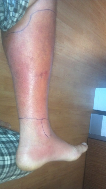
cellulitis
Pshawnoah / CC BY-SA

There are few points that must be kept in the back of head, that will help us to differentiate these two entities.

1. Overlying skin findings:

In osteomyelitis, the overlying soft tissues may appear normal or may be warm, mildly swollen, and occasionally erythematous, but in contrast to cellulitis, these surface findings are often sublte and induration in unusual. Spasm of overlying muscle is often intense, adding to discomfort and the adjacent joint may be held in flexion.

With osteomyelitis, swelling is usually more circumferential and the margins of the erythema less distinct. Cellulitis is more likely to occur in the area of a local wound, whereas no wound is typically present in osteomyelitis, because the mechanism is usually hematogenous seeding.

2. Tenderness:

Tenderness out of proportion to soft-tissue findings suggests osteomyelitis rather than soft-tissue infection or cellulitis.

3. Joint proximity:

Children with a limp or who are not weight bearing and have erythema close to a joint may have underlying osteomyelitis or septic arthritis.

4. Location of pain:

In cellulitis, the pain is localized only in the region affected, but in osteomyelitis the pain is circumferential, all around the affected bone.

5. Clinical improvement:

If the cellulitis of an extremity does not show significant improvement within 24 hours of antimicrobial therapy, a presumptive diagnosis of osteomyelitis should be made, and bone aspiration or open drainage should be performed.

6. X-rays:

Radiographs show the inflammatory response of the infection. In the early phase of the infection, soft tissue swelling is the only radiological finding. This sign may be overlooked or neglected. In osteomyelitis there is swelling of the deep soft tissue next to the bone, while in cellulitis, the superficial soft tissue is swollen.

References:

  1. Zitelli and Davis’ Atlas of Pediatric Physical Diagnosis – By Basil J. Zitelli, Sara C McIntire, Andrew J Nowalk
  2. David K. Hong, Kathleen Gutierrez, in Principles and Practice of Pediatric Infectious Diseases (Fifth Edition), 2018
  3. Osteomyelitis and septic arthritis in children by Hugo DE BOECK (Acta Orthop. Belg., 2005, 71, 505-515)
  4. The Sydney Children’s Hospital Network – Guideline: Paediatric Limb Cellulitis on Home Intravenous Therapy: Patient Management
  5. Moffet’s Pediatric Infectious Diseases: A Problem-oriented Approach By Randall G. Fisher, Thomas G. Boyce, Hugh L. Moffet

dr. sulabh kumar shrestha
Dr. Sulabh Kumar Shrestha, MS Orthopedics

He is the section editor of Orthopedics in Epomedicine. He searches for and share simpler ways to make complicated medical topics simple. He also loves writing poetry, listening and playing music. He is currently pursuing Fellowship in Hip, Pelvi-acetabulum and Arthroplasty at B&B Hospital.

2 shares
  • Facebook2
  • Twitter
Clinical Skills and Approaches DermatologyOrthopedics

Post navigation

Previous post
Next post

Related Posts

Clinical Skills and Approaches complications of transfusion

Perioperative Fluid Management

Jan 1, 2016May 5, 2016

Author: Sulabh Kumar Shrestha, KISTMCTH A) RELEVANT ANATOMY AND PHYSIOLOGY Details of the body fluid compartments are described here Microvessels for fluid exchange: The Exchange Vessels – capillaries and most proximal part of the venules Sinusoidal capillaries (liver, spleen, bone marrow): freely permeable to all solutes Fenestrated capillaries (glands, glomeruli, GIT):…

Read More
Clinical Skills and Approaches radial nerve course branches

Radial Nerve Anatomy : Course and Innervation

May 20, 2014Jun 1, 2024

Synonyms: Nervus radialis, Musculospiral nerve Recommended reading: Course 1. Origin: Terminal branch of Posterior cord of brachial plexus (Root value: C5, C6, C7, C8, T1) 2. Posterior axilla: It lies behind the axillary and upper part of the brachial arteries, passing anterior to tendons of subscapularis, lattisimus dorsi and teres…

Read More
Clinical Skills and Approaches

Drug Eruptions – When the Solution becomes Problem

Jun 18, 2020Jun 18, 2020

Before we start to develop our understanding of the drug eruptions. Let’s first lay down the foundation by clearing some basic concepts. What is an Allergy? What is meant by an adverse drug reaction? What is the diversity of allergic reactions due to drugs? Allergy This is a hypersensitivity disorder…

Read More

Leave a Reply Cancel reply

Your email address will not be published. Required fields are marked *

This site uses Akismet to reduce spam. Learn how your comment data is processed.

Shrestha SK. Cellulitis vs Early Osteomyelitis – Clinical features [Internet]. Epomedicine; 2020 Feb 22 [cited 2026 Jul 28]. Available from: https://epomedicine.com/clinical-medicine/cellulitis-vs-osteomyelitis/.

Pre-clinical (Basic Sciences)

Anatomy

Biochemistry

Community medicine (PSM)

Embryology

Microbiology

Pathology

Pharmacology

Physiology

Clinical Sciences

Anesthesia

Dermatology

Emergency medicine

Forensic

Internal medicine

Gynecology & Obstetrics

Oncology

Ophthalmology

Orthopedics

Otorhinolaryngology (ENT)

Pediatrics

Psychiatry

Radiology

Surgery

RSS Ask Epomedicine

  • What to study for Clinical examination in Orthopedics?
  • What is the mechanism of AVNRT?

Epomedicine weekly

  • About Epomedicine
  • Contact Us
  • Author Guidelines
  • Submit Article
  • Editorial Board
  • USMLE
  • MRCS
  • Thesis
©2026 Epomedicine | WordPress Theme by SuperbThemes