Vertebral Artery
I use the analogy of hand to remember the vertebral artery and it’s branches:
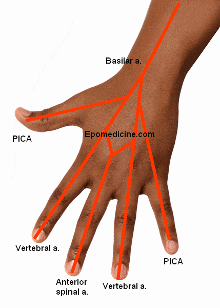
Origin: Branch of subclavian arteries
Course:
- Ascends through transverse foramina on C6 through C1 and enters posterior fossa through foramen magnum
- Continue up the ventral surface of medulla
- Converge at the ponto-medullary junction to form single basilar artery
- Branches are given inside cranial vault, once it has entered through foramen magnum
Supplies: Spinal cord, Medulla and Inferior cerebellum
Branches:
1. Anterior spinal artery (Single artery):
- Run down the front of the spinal cord
- Supplies ventrolateral 2/3rd of cervical spinal cord and ventrolateral medulla
2. Posterior spinal arteries:
- Bilaterally run down dorsolateral to spinal cord
- Supplies posterior 1/3rd of cervical spinal cord and posterior medulla
Clinical Correlate:
1. 10 Medullary arteries arising from segmental branches of aorta feeds anterior and posterior spinal artery along their course. In lower thoracic/upper lumbar region, Large segmental artery exists and usually on Left side – named as Adamkiewicz. It’s injury can result in paraplegia due to its lower thoraci/upper lumbar location.
2. Anterior spinal infarct can only affect the arm fibers of the lateral corticospinal tracts without affecting the leg fibers and vice versa (a posterior spinal artery infarct can affect the leg and not the arm fibers), because the border of the anterior and posterior spinal arterial territories lies within the corticospinal tract systems in the lateral funiculi.
3. Occlusion of vertebral artery or Anterior spinal artery can result in Medial medullary or Djerine’s syndrome.
3. Posterior Inferior Cerebellar Artery (PICA):
- Supplies all of medulla except antero-median part
- Supplies all of the inferior cerebellum and medial part of middle cerebellum
Clinical Correlate: PICA injury leads to PICA or Wallenberg Syndrome
4. Meningeal branch:
- Supplies falx cerebri
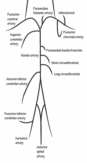
Basilar Artery
Origin: Joining of 2 vertebral arteries at ponto-medullary junction
Course:
- Ascends along the midline of pons
- Terminates near rostral border of pons by dividing into 2 Posterior cerebral arteries
Supplies: Pons, Anteroinferior and superior cerebellum and Inner ear
Clinical correlate: Obstruction of basilar artery damaging the bilateral ventral pons give rise to Locked-in Syndrome. Because the tegmentum of the pons is spared, the patient has a spared level of consciousness, preserved vertical eye movements, and blinking. The corticospinal and corticonuclear tracts are affected bilaterally. The oculomotor and trochlear nerves are not injured. Patients are conscious and may communicate through vertical eye movements.
Branches:
Since, there is Posterior Inferior Cerebellar Artery – there must also be Anterior Inferior Cerebellar Artery (AICA) and Superior Cerebellar Artery (SCA). The branches from down to up are:
1. AICA:
- Supplies dorsolateral part of caudal pons and antero-inferior region of cerebellum
- Gives rise to labyrinthine artery in 85% cases
Clinical correlate:
Occlusion of AICA can result in Lateral Pontine Syndrome or Marie-Foix syndrome. It is similar to Lateral medullary syndrome but can be localized by lesions of CN VII, CN VIII and other nucleus of CN V except spinal nucleus of CN V which is also injured in medullary syndromes. AICA occlusion is more specifically localized by presence of CN VII and CN VIII lesions as it is present in the caudal pons.
2. Labyrinthine artery:
- Usually originates from AICA but can originate from the basilar artery
- Follows the course of CN VIII and supplies the internal ear
3. Pontine branches:
- Supplies anterior and lateral part of pons through paramedian and circumferential branches
Clinical correlate:
Occlusion of paramedian branches of basial artery results in Medial pontine syndrome (Foville syndrome). This is similar to medial medullary syndrome but can be localized by the findings of CN VI (medial strabismus due to lateral rectus paralysis and lateral gaze paralysis if PPRF is involved) and VII lesions (LMN type of facial palsy).
Occlusion of the paramedian and circumferential branches can result in Ventral pontine syndrome (Millard-Gubler Syndrome). It presents with contralateral limb weakness (corticospinal tract involvement) and ipsilateral CN VI and VII defects.
4. Superior cerebellar artery (SCA):
- Supplies dorsolateral part of rostral pons and caudal midbrain
- Supplies superior cerebellum and lateral region of mid-cerebellum
Clinical correlate:
Occlusion of SCA can result in Lateral Pontine Syndrome or Marie-Foix syndrome. It is similar to Lateral medullary syndrome but can be localized by lesions of CN VII, CN VIII and other nucleus of CN V except spinal nucleus of CN V which is also injured in medullary syndromes. SCA occlusion is more specifically localized by presence of CN V lesions as it is present in the rostral pons.
5. Posterior cerebral arteries:
- Paramedian and circumferential branches supply the midbrain
- Forms circle of willis to supply brain
Clinical correlate:
Occlusion of posterior cerebellar arteries can result in Medial midbrain syndrome (Weber syndrome). It is characterized by contralateral limb weakness (corticospinal tract involvement), contralateral lower facial weakness (corticobulbar fiber involvement i.e. UMN type of facial palsy) and ipsilateral CN III lesion.
Parinaud syndrome (Dorsal midbrain syndrome): This is caused due to pineal tumor compressing the superior colliculi. It compresses the vertical gaze center at the rostral interstitial nucleus of medial longitudinal fasciculus (riMLF) leading to vertical gaze palsy. It is accompanied by bilateral pupillary abnormalities and signs of elevated ICP (cerebral aqueduct compression).
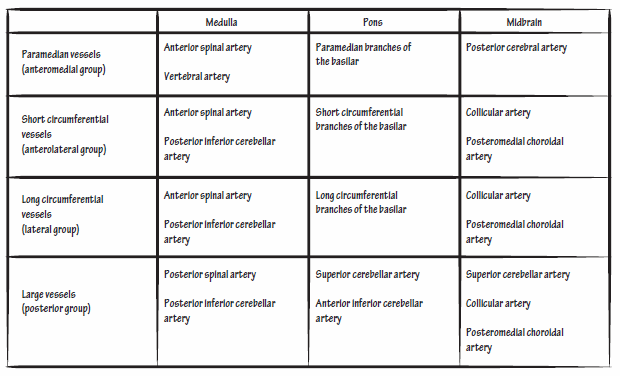
Arterial Supply of Brainstem