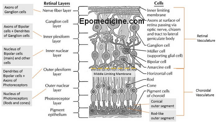
The ten layers of retina – this microscopic anatomy is frequently asked in examinations and also important from the physiological viewpoint. There are plenty of mnemonics around the web, but we will proceed in a different approach to remember the 10 retinal layers easily.
A. Retina is 3 neuron system composed of 3 layers of cells:
From out to in –
- PhotoReceptor layer
- Pigmented epithelium with Zona occludens (outer blood-retina barrier)
- Photoreceptors
- Outer limiting membrane
- Outer nuclear layer
- Outer plexiform later
- Bipolar cell layer (1st order neuron)
- Inner nuclear layer
- Ganglion cell layer (2nd order neuron)
- Inner plexiform layer
- Ganglion cell layer
- Nerve fiber layer (Axons of ganglion cell layer that form optic nerve)
- Inner limiting membrane
Remember: RBG (Red, Blue, Green) – the color model for color vision
Where are the 3rd order neurons? In the Lateral Geniculate Body (LGB).
B. Orientation:
- Inner – refers to layers which are close to the vitreous humor
- Outer – refers to layers which are close to the choroid
C. Nuclei of Retinal cells:
These nuclei are arranged in a layer known as Nuclear layer.
- Nuclei of Photoreceptors – forms Outer Nuclear Layer
- Nuclei of Bipolar cells (1st order neuron), Horizontal cells (outer to Bipolar cells), Amacrine cells (inner to Bipolar cells) and Muller cell (supporting glial cells) – forms Inner Nuclear Layer
Horizontal cells: help sharpen the receptive field of bipolar cells
Amacrine cells: help sharpen the response of ganglion cells
D. Axons and Dendrites of Retinal cells:
These axons and dendrites (synapses) are arranged in a layer known as Plexiform layer.
Plexiform layer lies just inside the corresponding Nuclear layer:
- Outer plexiform layer (OPL): Between outer nuclear layer and inner nuclear layer
- Synapses between Photoreceptors and Bipolar cells forms Middle Limiting Membrane (MLM).
- Retina external to MLM is avascular (dependent upon choroidal vasculature) and Internal to MLM is vascular (Central retinal artery).
- Hence, outer plexiform layer is a watershed zone between dual vascular supply – role in localization of edema fluid and hard exudates in this layer.
- Inner plexiform layer (IPL): Between inner nuclear layer and ganglion cell layer
- Synapses between Bipolar cells, Ganglion cells and Amacrine cells.
E. Axons of Ganglion Cells form Nerve Fiber Layer:
- Which go on to Optic nerve (CN II)
F. Inner Limiting Membrane:
- The only true basement membrane.
- Synthesized by foot process of Muller cells.
G. Shape of Photoreceptors:
The name comes from the outer segment of these receptors, which contains photopsin:
- Rods: Outer segment is Rod-like cylindrical
- Cones: Outer segment is Conical
Rods: Dark vision and Motion – peak sensitivity to light at wavelength 505 nm.
Cones: Light vision, Sharp vision and Color discrimination
- 3 types: Red (565 nm), Blue (535 nm) and Green (440 nm)
With this Knowledge, now lets draw the microanatomy of retina:
Schematic Drawing of Retinal Layers
Each retina has 100 million rods, 3 million cones and only 1.6 million ganglion cells. Thus 60 rods and 2 cones converge on each ganglionic cell and optic nerve fiber. In central retina (fovea) fewer rods and cones converge on each fiber progressively increasing the acuity of vision. Central fovea (foveola) has 35,000 slender cones and no rods converging on almos exactly equal number of nerve fibers leading to maximum color vision acuity. Peripheral retina has much greater sensitivity to weak light because rods are 30-300 times more sensitive to light than cones and ~200 rods converge on a single optic nerve fiber in peripheral portion of retina.
Lateral inhibition (by horizontal and amacrine cells) provide contrast detection and enhancement.
Rod cells in Light and Dark
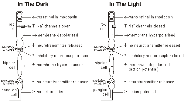
- Rods – Bipolar cells synapse = Inhibitory synapse
- Bipolar cells – Ganglion cells synapse = Excitatory synapse
- Hence, to generate action potential in Ganglion cells: Rod cells must be inhibited (Inhibtion of inhibitory fibers)
- There are cGMP gated Na+ channels on rods.
- cGMP is abundant in dark and decreased in light.
- Depolarization of rods occurs in dark and repolarization in light.
The process of phototransduction (response to light) in rods and cones is virtually the same. Unlike other receptors, the photoreceptors are hyperpolarized (instead of depolarized) and donot porduce action potential.
The process of phototransduction: